

If your pathology report says epithelioid mesothelioma—or you are researching the diagnosis for someone in your family—this page is a place to start. Epithelioid is the most common histologic subtype, accounting for about 6 in 10 mesotheliomas overall, although the reported proportion varies by anatomical site, study population, and specimen type.1 Compared with biphasic and sarcomatoid disease, it is generally associated with longer survival and is the subtype most often considered for the full range of treatment options. Histology is important, but it is only one part of the picture.
Evidence scope. Mesothelioma can arise in the pleura, peritoneum, pericardium, or tunica vaginalis—the membrane around a testicle. The same broad histologic subtypes are recognized across these sites, but the grading, prognosis, and treatment evidence attached to them is not necessarily interchangeable. Most numerical outcomes and clinical-trial results discussed on this page come from diffuse pleural mesothelioma. Peritoneal mesothelioma has a smaller, separate evidence base and different treatment pathways. Primary pericardial mesothelioma and mesothelioma of the tunica vaginalis are exceptionally rare, so the available evidence comes mainly from registries, small series, and case reports. Unless stated otherwise, statistics on this page refer to pleural disease.2–4
Epithelioid is the most favorable histology on average, not a promise about one person's outcome. In a national cancer-registry (SEER) study of 1,183 patients with stage I–III pleural mesothelioma diagnosed from 2004 through 2010, before immunotherapy became routine, median overall survival was 14 months for epithelioid disease, 10 months for biphasic disease, and 4 months for sarcomatoid disease.5 Because the study excluded stage IV disease, its medians are likely higher than they would be for all patients. They are historical group medians, not a forecast for an individual patient. Stage, tumor grade, lymph-node involvement, age, overall health, anatomical site, and treatment all matter, and outcomes in the modern treatment era may differ from older registry data.
You do not need to be an expert in pathology to make sense of your report. Most patients arrive here with a diagnosis they did not choose, a vocabulary they do not speak, and questions that have not been fully answered. The purpose of this page is to help you understand what your report says, what the findings can—and cannot—tell you about treatment and prognosis, and what questions may be worth raising with your medical team.
You don't need the product names or old records. No fee unless we recover compensation for your family.
Quick answers
| What does “epithelioid” mean in my pathology report? | It describes how the tumor cells look and grow under a microscope. Epithelioid cells are usually cohesive and polygonal, cuboidal, or columnar, and they may form tubules, papillae, trabeculae, or solid sheets. Epithelioid is one of the three main mesothelioma histologic subtypes; the other two are sarcomatoid and biphasic. |
|---|---|
| Is epithelioid the “good” mesothelioma subtype? | No mesothelioma subtype is “good.” Epithelioid is generally the most favorable of the three because it is associated, on average, with longer survival and a broader range of treatment considerations. An individual outlook still depends on location, stage, grade, lymph-node involvement, overall health, and treatment. |
| What is WHO grading, and why does it matter? | For diffuse pleural epithelioid mesothelioma, the pathologist first assigns a three-tier nuclear grade from nuclear atypia and mitotic activity. That nuclear grade is then combined with the presence or absence of tumor necrosis to produce the final two-tier WHO tumor grade: low or high. Grade provides prognostic information, but it is not a stand-alone treatment rule. |
| Do the survival numbers on this page apply to peritoneal, pericardial, or tunica-vaginalis mesothelioma? | Not automatically. Most published grading, survival, and clinical-trial data concern pleural disease. Peritoneal mesothelioma has different treatment pathways and its own outcome literature. Pericardial and tunica-vaginalis mesotheliomas are so rare that the evidence is much more limited. |
| Am I a candidate for surgery with epithelioid mesothelioma? | Epithelioid histology may make a surgical evaluation more likely than non-epithelioid histology, but it does not make surgery appropriate by itself. Candidacy depends on whether the disease is technically resectable, stage and lymph-node status, cardiopulmonary reserve, performance status, other medical conditions, the patient's goals, and review by an experienced multidisciplinary team. |
| Does the epithelioid subtype change my legal options? | Usually not by itself. Eligibility for a lawsuit, asbestos trust claim, or benefit program depends on the diagnosis, exposure evidence, responsible companies or worksites, program rules, jurisdiction, and filing deadlines. Histology may affect medical proof or case planning, but it does not guarantee that every recovery channel applies. |
| How long do I have to file a claim? | It depends on the applicable state law, the type of claim, and whether the matter involves personal injury or wrongful death. Some deadlines are short. Prompt review is the safest way to determine which deadlines apply. |
| Can a spouse or adult child call for the patient? | Yes. A spouse, adult child, or other family member can ask questions and begin gathering information. A power of attorney is not ordinarily needed simply to make an initial inquiry, although formal authority may be required for later decisions or filings. |
“I started this firm after learning that the cancers that killed my father and grandparents were caused by asbestos, something we did not realize until it was too late to hold anyone accountable. That experience is why we built this site: so that no other family misses their chance to ask the questions, hear the options, and decide for themselves what to do.”
What epithelioid mesothelioma is
The term epithelioid describes the appearance and arrangement of the tumor cells under a microscope. The cells are often polygonal, cuboidal, or columnar and tend to remain cohesive. They may grow in tubulopapillary, trabecular, adenomatoid, microcystic, solid, or micropapillary patterns. These epithelial-like shapes and structures help a pathologist classify the tumor, but appearance alone is not enough to establish a mesothelioma diagnosis. The microscopic findings must be interpreted with immunohistochemistry, evidence of invasion when available, molecular tests when appropriate, imaging, and the clinical setting.2,3
At the population level, epithelioid histology is associated with better outcomes than biphasic or sarcomatoid histology.5 That does not mean every epithelioid tumor remains localized, can be removed surgically, or will respond to a particular treatment. Histologic subtype does not tell doctors how far the cancer has spread, and it does not replace staging, assessment of lymph nodes, or evaluation of the patient's overall condition.
Mesothelioma most often begins in the pleura, the membrane lining the lungs and inside of the chest wall. It can also arise in the peritoneum, the lining of the abdominal cavity, and much more rarely in the pericardium or tunica vaginalis. The broad histologic label may be the same at each site, but its prognostic and treatment implications must be interpreted in the context of the anatomical location. A pleural survival estimate or treatment trial should not be applied automatically to a peritoneal or rare-site tumor.3,4
How epithelioid pathology varies
The 2021 WHO classification recognizes meaningful variation within epithelioid diffuse pleural mesothelioma. Pathologists are encouraged to report architectural patterns, cytologic and stromal features, necrosis, and tumor grade because these findings can add prognostic information.2,3 They describe associations seen across patient groups; no single pattern or grade determines an individual outcome or automatically selects a treatment.
A biopsy also samples only part of a tumor. Architectural proportions, necrosis, and even subtype can differ between a biopsy and a later resection specimen, so pathology findings should always be interpreted in light of specimen size and sampling.6
Architectural patterns
Epithelioid mesothelioma can contain one pattern or a mixture of several:
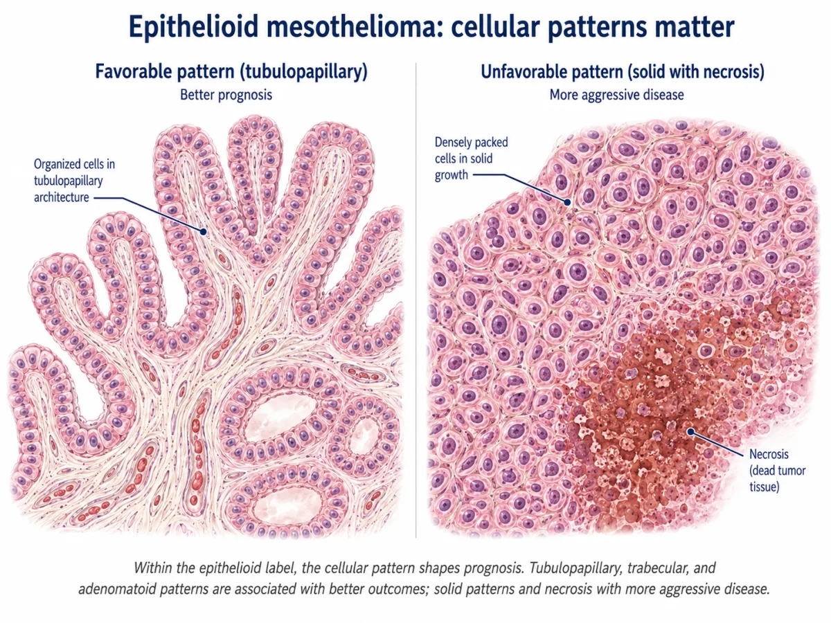
- Patterns associated with more favorable outcomes include tubulopapillary, trabecular, and adenomatoid architecture.
- Patterns associated with less favorable outcomes include micropapillary architecture and a solid pattern when solid growth makes up more than 50 percent of the sampled tumor.
- Other recognized patterns, including microcystic growth, may be reported even when their independent prognostic meaning is less clearly established.2,3
The amount of each pattern can matter, but the percentage should not be used as a stand-alone treatment cutoff. A small biopsy may not reproduce the proportions found in a larger specimen.
Cytologic and stromal features
A pathology report may also describe features of the individual cells or the tissue surrounding them:
- Predominant myxoid stroma—myxoid material in more than 50 percent of the tumor, with less than 50 percent solid growth—has been associated with a more favorable prognosis.
- Rhabdoid or pleomorphic cytologic features have been associated with more aggressive behavior and should be reported when present.
- Tumor necrosis, meaning areas of dead tumor tissue, is an unfavorable finding and is incorporated into the WHO grading framework described below.
- Lymphohistiocytoid morphology is rare and can occur in more than one main histologic subtype. Some small series have associated it with a more favorable outcome, but it should not be treated as an epithelioid-only feature or a definitive prognostic rule.2,3
These descriptors add detail to the main diagnosis. They do not create additional principal mesothelioma subtypes.
WHO grading for epithelioid diffuse pleural mesothelioma
The WHO grading framework uses a two-step process. It is important to distinguish the three-tier nuclear grade from the final two-tier tumor grade.2,7
First, the pathologist scores nuclear atypia and mitotic activity:
| Component | Score 1 | Score 2 | Score 3 |
|---|---|---|---|
| Nuclear atypia | Mild | Moderate | Severe |
| Mitotic count per 2 mm² | 0–1 | 2–4 | 5 or more |
The two scores are added:
- Total score 2–3 = nuclear grade 1
- Total score 4–5 = nuclear grade 2
- Total score 6 = nuclear grade 3
The nuclear grade is then combined with necrosis to produce the final WHO tumor grade:
| Final WHO tumor grade | Criteria |
|---|---|
| Low grade | Nuclear grade 1, with or without necrosis; or nuclear grade 2 without necrosis |
| High grade | Nuclear grade 2 with necrosis; or nuclear grade 3, with or without necrosis |
This is a two-tier low-grade/high-grade system, not a four-grade WHO system. The research that informed the framework identified several combinations of nuclear grade and necrosis with different median outcomes, but those study-specific survival groups are not four official WHO grades and should not be used as fixed predictions for an individual patient.2,7
Grading should be based on the area showing the highest-grade features. It can be reported on biopsy and resection specimens, but a limited biopsy may not capture the highest-grade portion of a heterogeneous tumor.2,3,6 If an epithelioid pleural mesothelioma report does not address nuclear atypia, mitotic activity, necrosis, or final tumor grade, it is reasonable to ask whether the tissue can be reviewed by a pathologist with mesothelioma expertise.
Molecular markers
BAP1, MTAP, and CDKN2A are commonly used as ancillary diagnostic markers. Their best-established role is helping a pathologist distinguish a malignant mesothelial proliferation from a benign or reactive one. They are interpreted only after the cells have been shown to be mesothelial; by themselves, these tests do not distinguish mesothelioma from every other cancer, determine the anatomical stage, or select a standard treatment.2,8–10
| Marker | How it is usually tested | What an abnormal result can support | Important limitation |
|---|---|---|---|
| BAP1 | Immunohistochemistry looks for loss of nuclear BAP1 protein in tumor cells. | BAP1 loss strongly supports malignancy in a mesothelial proliferation and is particularly useful in epithelioid and biphasic tumors. | Many mesotheliomas retain BAP1, so a normal result does not rule out the disease. Tumor BAP1 loss does not by itself show that an alteration was inherited. |
| MTAP | Immunohistochemistry looks for loss of cytoplasmic MTAP protein. | MTAP loss can support malignancy and often acts as a practical surrogate for deletion of the nearby CDKN2A region on chromosome 9p21. | The surrogate is not perfect and has been validated mainly in pleural mesothelioma; retained MTAP does not rule out mesothelioma or a CDKN2A alteration. Results must be interpreted with the morphology and other tests. |
| CDKN2A | Fluorescence in situ hybridization (FISH) tests for homozygous deletion of the gene. | Homozygous CDKN2A deletion strongly supports malignancy in a mesothelial proliferation and can be especially helpful when BAP1 is retained. | Not every mesothelioma has the deletion. A negative FISH result does not exclude malignancy. |
A BAP1 abnormality found only in tumor tissue should be treated as a tumor finding; it does not establish whether the alteration is inherited. Germline status requires separate testing, usually on blood or saliva. The 2025 ASCO guideline recommends offering germline genetic testing to all patients with mesothelioma.11
An inherited predisposition and asbestos exposure can coexist. Neither a tumor BAP1 result nor a germline result, standing alone, establishes or excludes the role of asbestos in an individual case. That question requires the exposure history and other case-specific medical and factual evidence.
Broader molecular profiling may help identify eligibility for a clinical trial, but BAP1 loss, MTAP loss, or CDKN2A deletion does not by itself imply that an approved targeted therapy will be effective.
How epithelioid mesothelioma is diagnosed and classified
Diagnosing epithelioid mesothelioma is an integrated process, not a matter of finding one “positive” stain. The pathologist considers the amount and depth of tissue, the microscopic appearance of the cells, evidence of invasion when it can be assessed, immunohistochemistry to establish mesothelial lineage and exclude look-alike cancers, ancillary tests that can support malignancy, and the clinical and imaging findings. Once mesothelioma is established, the pathologist classifies the histologic subtype and, for diffuse pleural epithelioid mesothelioma, reports prognostic features and WHO tumor grade when the specimen permits.2,3,11,12
The sampling methods described below principally concern pleural mesothelioma. A tumor arising in the peritoneum, pericardium, or tunica vaginalis may require a different biopsy route and a site-specific diagnostic approach.
Tissue sampling
For a patient with suspected pleural mesothelioma who is being evaluated for anticancer treatment, thoracoscopic biopsy is generally preferred when it is medically feasible. A camera is passed through the chest wall so the pleural surfaces can be inspected and several tissue samples can be taken from representative areas. This approach usually provides more tissue—and a better opportunity to assess invasion, subtype, architecture, necrosis, and grade—than a small superficial sample. When thoracoscopy is not feasible, an image-guided core needle biopsy of an accessible pleural mass or area of thickening may be appropriate.11,12
Pleural-fluid cytology can sometimes contribute to, or in selected cases establish, the diagnosis when the cellular and ancillary findings are convincing. It also has important limitations. A negative or indeterminate fluid result does not rule out mesothelioma, and fluid alone often cannot show stromal invasion or provide enough representative tissue to determine subtype, architectural proportions, necrosis, or grade with confidence.12
Sampling error matters because mesothelioma can be heterogeneous. A superficial biopsy may contain reactive mesothelial cells or fibrosis while missing the invasive tumor beneath it. A small biopsy may establish mesothelioma but fail to capture the tumor’s highest-grade area or a second histologic component. Conversely, a limited sample can make a feature appear more dominant than it is in the tumor as a whole.6,12
If the first specimen is inconclusive or does not contain enough tissue for clinically important classification, the next step is not automatically another procedure. Existing slides and tissue blocks can often be reviewed first by a pathologist with mesothelioma expertise, who may recommend additional stains or determine whether more tissue is truly needed. When further biopsy is proposed, the potential diagnostic benefit should be weighed against the risks and the possibility that treatment should not be delayed.
Immunohistochemistry: a panel, not a single definitive stain
Immunohistochemistry, usually abbreviated IHC, uses antibodies to detect proteins in or on cells. No single IHC marker is a “gold standard” for epithelioid mesothelioma, and no stain should be interpreted by itself. Pathologists ordinarily use a panel selected for the specific differential diagnosis—commonly at least two mesothelial-lineage markers and at least two carcinoma markers, with additional organ-specific stains when needed.12,13
| Diagnostic question | Commonly used markers | What the result can show |
|---|---|---|
| Do the tumor cells show mesothelial differentiation? | Calretinin, WT1, D2-40 (podoplanin), and cytokeratin 5/6 | A compatible pattern supports mesothelial lineage. Individual markers vary in sensitivity and specificity, so one positive stain is not enough. |
| Could the tumor instead be a carcinoma involving the pleura? | Claudin-4, MOC-31, Ber-EP4, and sometimes CEA | Positive staining—particularly convincing membranous claudin-4—favors carcinoma. A negative result alone does not prove mesothelioma. |
| Where might a carcinoma have originated? | Markers chosen for the clinical differential, such as TTF-1 and Napsin A for lung adenocarcinoma, GATA3 or mammaglobin for breast origin, PAX8 for renal or Müllerian origin, or other site-directed stains | These help identify or exclude a likely primary site. The exact panel depends on the patient’s history, imaging, sex, and tumor location. |
Calretinin, WT1, D2-40, and cytokeratin 5/6 are useful mesothelial markers, but none is invariably positive in every epithelioid mesothelioma and none is completely specific in every diagnostic setting. Likewise, MOC-31 and Ber-EP4 can occasionally stain mesothelioma, which is one reason claudin-4 has become a particularly useful broad carcinoma marker. The pathologist interprets the pattern, intensity, location, and combination of staining rather than treating each stain as a yes-or-no answer.12,13
An important distinction follows: a mesothelial-positive and carcinoma-negative IHC pattern can establish that the cells are mesothelial, but it does not by itself prove that the proliferation is malignant. Reactive mesothelial cells can express the same lineage markers. Malignancy is established through invasion when demonstrable and, in appropriate cases, through the ancillary tests described next.
Ancillary tests that help establish malignancy
The most commonly used malignancy-associated tests are BAP1 immunohistochemistry, MTAP immunohistochemistry, and CDKN2A fluorescence in situ hybridization, or FISH. BAP1 and MTAP are protein stains rather than genetic sequencing tests; CDKN2A FISH looks for homozygous deletion of the 9p21 region.8–10,12,13
- Loss of nuclear BAP1 expression in a mesothelial proliferation strongly supports malignancy.
- Loss of cytoplasmic MTAP expression also supports malignancy and often serves as a practical surrogate for deletion of the nearby CDKN2A region.
- Homozygous CDKN2A deletion by FISH strongly supports malignancy and can be especially useful when BAP1 is retained.
These tests are highly useful when abnormal, but they are not perfectly sensitive. Retained BAP1 or MTAP and a negative CDKN2A FISH result do not rule out mesothelioma. The results must be interpreted with the morphology, IHC lineage panel, specimen type, imaging, and clinical history. Tumor BAP1 loss also does not establish that a BAP1 alteration was inherited; germline status requires separate testing, as discussed earlier on this page.
Broader tumor sequencing may be ordered to identify a clinical-trial opportunity or another actionable finding. It is not a substitute for the core pathology workup, and BAP1, MTAP, or CDKN2A findings do not by themselves select one standard mesothelioma treatment.
Subtype and WHO grade
After the pathologist establishes mesothelioma, the tumor should be classified as epithelioid, sarcomatoid, or biphasic whenever the tissue permits. For diffuse pleural epithelioid mesothelioma, current reporting guidance also calls for architectural patterns, relevant cytologic and stromal features, necrosis, and the final WHO tumor grade.2,3,11,12
As explained in the preceding section, WHO grading is a two-step process. Nuclear atypia and mitotic activity produce a three-tier nuclear grade; nuclear grade is then combined with necrosis to produce the final two-tier tumor grade: low or high. The report may list the component scores as well as the final grade.
A few limitations are worth keeping in mind:
- Grade is based on the highest-grade area identified, but a limited biopsy may not contain that area.
- A report that omits grade does not mean the tumor is low grade. It may mean the specimen was insufficient, the report predates current guidance, or grading was not performed.
- Grade provides prognostic information, but it is not a stand-alone rule for surgery, chemotherapy, or immunotherapy. Stage, resectability, lymph-node status, overall health, and the patient’s goals remain central to treatment planning.
If a pleural epithelioid report does not address subtype confidence, necrosis, mitotic activity, nuclear atypia, or final tumor grade, it is reasonable to ask whether the available tissue is adequate for those assessments and whether specialist review would add useful information.
When expert pathology review is important
Mesothelioma is uncommon and can resemble reactive mesothelial proliferations, metastatic adenocarcinoma, serous carcinoma, squamous carcinoma, and other tumors. A second review by a pathologist with mesothelioma expertise is particularly reasonable when:
- The diagnosis is uncertain or the report uses language such as “atypical mesothelial proliferation,” “suspicious,” or “cannot exclude mesothelioma.”
- The sample was small, superficial, crushed, or largely fibrotic.
- The IHC panel was limited or the stains do not form a coherent pattern.
- The report does not clearly state the histologic subtype or, for pleural epithelioid disease, does not address grade when adequate tissue appears to be available.
- The pathology does not fit the imaging or clinical course.
- An irreversible or high-risk decision—particularly cytoreductive surgery—is being considered.
A second opinion usually does not require the patient to travel. The original laboratory can send stained slides, unstained slides, and tissue blocks to the consulting center, which then issues a separate report. A new biopsy may still be necessary in some cases, but specialist review of the existing material is often the appropriate first step.
Want help organizing the questions raised by your pathology report? A member of our litigation team—not a medical screener—can review the document with you for case-intake purposes, help identify where terms such as subtype, grade, BAP1, MTAP, and CDKN2A appear, and help you prepare questions for your medical team. We do not diagnose disease or recommend treatment. The conversation is free and creates no obligation.
Call 833-4-ASBESTOS or request a free case review.
What epithelioid histology means for treatment
Unless stated otherwise, this section concerns diffuse pleural mesothelioma. Peritoneal mesothelioma follows a different treatment pathway, often including evaluation for cytoreductive surgery and heated intraperitoneal chemotherapy, or HIPEC.14 See our peritoneal mesothelioma page for that site-specific discussion.
Epithelioid histology generally keeps the widest range of pleural treatment options in consideration, but it does not choose a treatment. Stage, lymph-node involvement, resectability, performance status, cardiopulmonary reserve, other medical conditions, prior treatment, and the patient's goals remain central. The WHO tumor grade refines prognosis; it is not a validated stand-alone rule for choosing surgery, chemotherapy, or immunotherapy.11
Surgery may be discussed for a narrow, highly selected group
Epithelioid histology is generally a prerequisite for modern cytoreductive-surgery consideration, not proof that surgery will benefit a particular patient. In the randomized MARS2 trial, extended pleurectomy/decortication plus chemotherapy was associated with worse survival over two years (median 19.3 versus 24.8 months), more serious adverse events, and worse quality of life than chemotherapy alone. Most MARS2 participants had epithelioid disease. Current ASCO guidance therefore limits surgical consideration to highly selected, comprehensively staged patients with early-stage epithelioid disease and favorable characteristics at experienced centers, preferably within a clinical trial.11,15
Procedures performed primarily to control pleural fluid, improve breathing, or relieve pain are different from radical cytoreductive surgery and may remain appropriate when tumor-removing surgery is not recommended.
Unresectable pleural disease has several systemic options
For previously untreated unresectable pleural disease, recognized first-line options include platinum-pemetrexed chemotherapy, with bevacizumab for selected patients; nivolumab plus ipilimumab; and pembrolizumab plus platinum-pemetrexed chemotherapy. No randomized trial directly compares all three approaches, and no one regimen is automatically best for every patient with epithelioid disease.11
In CheckMate 743, the average relative survival advantage of nivolumab plus ipilimumab over chemotherapy was smaller and statistically less certain in the epithelioid subgroup than in the pooled non-epithelioid subgroup. A minority of patients with epithelioid disease nevertheless achieved durable multi-year survival; the five-year findings are discussed in the prognosis section below.16
The choice among systemic approaches can depend on the need for relatively prompt tumor shrinkage, autoimmune disease or transplant history, kidney and hearing function, other medical conditions, toxicity preferences, treatment logistics, and the patient's goals. PD-L1 is not required for nivolumab-ipilimumab and has not been established as a sufficiently reliable stand-alone selection test. Tumor grade likewise does not automatically select a regimen.11
What the subtype does—and does not—decide
The practical effect of an epithelioid diagnosis is that surgery, chemotherapy, and immunotherapy may all enter the discussion more often than with other histologies. It does not mean that the tumor is resectable, that chemotherapy is always preferred, or that immunotherapy is unlikely to help.
A specialty review is particularly valuable when surgery is under consideration, the pathology is uncertain, major medical conditions complicate systemic treatment, or a clinical trial may be available. Much of the day-to-day treatment can often be delivered closer to home with a specialty center advising on the overall plan.
For details about pleurectomy/decortication and extrapleural pneumonectomy, the MARS2 debate, chemotherapy and immunotherapy regimens, adverse effects, radiation, tumor treating fields, later-line treatment, clinical trials, and costs, see our pleural mesothelioma treatment page.
Prognosis and outcomes for epithelioid mesothelioma
Epithelioid mesothelioma is associated with the longest survival of the three principal histologic subtypes on average. That is useful information, but it is not an expiration date or a prediction of what will happen to one person. Prognosis depends on the anatomical site, stage, lymph-node involvement, tumor grade, the patient's overall condition, the treatments that are medically appropriate, and how the cancer responds.
As explained at the beginning of this page, most of the numerical evidence below concerns diffuse pleural mesothelioma. Peritoneal mesothelioma follows a different treatment pathway, and outcomes from pericardial or tunica-vaginalis mesothelioma are too sparsely reported to support the same kind of estimates.
What historical population data can—and cannot—tell you
In the SEER analysis of 1,183 patients with stage I–III pleural mesothelioma diagnosed from 2004 through 2010, median overall survival was 14 months for epithelioid disease, compared with 10 months for biphasic disease and 4 months for sarcomatoid disease.5 Among patients with epithelioid histology, the median was 19 months in the group that underwent cancer-directed surgery and 10 months in the group that did not.5
That surgical comparison was observational, not randomized. Patients selected for surgery were likely to differ from nonsurgical patients in age, stage, functional status, tumor extent, and other ways that also affect survival. The study therefore shows an association between surgical selection and longer survival; it does not prove that surgery itself produced the entire difference. The cases also came from a treatment era that predated routine checkpoint immunotherapy. These medians remain useful historical context, but they should not be presented as a current, individualized forecast.
Pleural and peritoneal outcomes should not be blended. Selected patients with diffuse peritoneal mesothelioma may be considered for cytoreductive surgery and hyperthermic intraperitoneal chemotherapy (HIPEC), a treatment pathway that does not have a direct pleural equivalent. In a multi-institutional registry of 405 patients treated with cytoreductive surgery (372 of whom also received HIPEC), median overall survival was 53 months and five-year survival was 47 percent; a later Australian single-center series of 81 patients reported the same 53-month median and 49 percent five-year survival.17,18 Those are retrospective surgical-series results—not general-population estimates or randomized comparisons—and should not be substituted for pleural survival figures or applied to every patient with peritoneal disease.
Tumor grade refines prognosis within epithelioid pleural disease
For diffuse pleural epithelioid mesothelioma, the final WHO tumor grade is low or high. Low-grade tumors are associated with more favorable outcomes than high-grade tumors across patient groups, but grade does not determine how long a particular patient will live and does not select a treatment by itself.2,7
The multi-institutional study that helped establish the grading framework reported four combinations of nuclear grade and necrosis with different median outcomes. These are study-specific prognostic strata, not four official WHO grades:
| Nuclear grade and necrosis in the study cohort | Median overall survival in that cohort |
|---|---|
| Nuclear grade 1 without necrosis | 29 months |
| Nuclear grade 1 with necrosis, or nuclear grade 2 without necrosis | 16 months |
| Nuclear grade 2 with necrosis | 10 months |
| Nuclear grade 3, with or without necrosis | 8 months |
The study included 776 patients treated at multiple institutions from 1998 through 2014. Its medians describe groups treated during that period; they are not fixed expectations for a patient receiving care today.7 The current WHO system combines the first two rows into low-grade disease and the last two into high-grade disease, as described earlier on this page.
A limited biopsy may not contain the tumor's highest-grade area or may not show necrosis that is present elsewhere. Grade should therefore be interpreted together with specimen size, staging, imaging, and the rest of the pathology findings.6
Factors that shape an individual prognosis
Several findings provide more context than the epithelioid label alone:
- Anatomical site. Pleural and peritoneal mesothelioma have different staging approaches, treatment pathways, and outcome literature. Rare-site tumors require especially cautious interpretation because published series are small.
- Stage, lymph nodes, and tumor extent. The amount and distribution of disease affect whether local treatment is technically possible and which systemic or symptom-directed options are appropriate.
- Final tumor grade and other pathology features. High grade, necrosis, micropapillary architecture, and predominant solid growth are associated with less favorable outcomes; lower grade and certain organized architectural patterns are associated with more favorable outcomes.2,7
- Performance status, organ function, and other medical conditions. These factors affect both prognosis and the ability to receive particular treatments. They also help explain why clinical-trial results may not reproduce exactly in a broader patient population.
- Treatment received and treatment response. A durable response can extend survival, but pathology cannot predict in advance which individual patient will achieve one.
- Quality and representativeness of the specimen. A small biopsy may correctly establish epithelioid mesothelioma while underrepresenting grade, necrosis, architectural proportions, or a second histologic component.6,12
A consultation at a center with substantial mesothelioma experience can be useful when the diagnosis is uncertain, surgery is being considered, or the systemic-treatment choices are difficult. That does not mean every part of care must occur far from home. Specialty review and local treatment can often be coordinated.
What modern treatment changes
Historical registry data cannot fully describe outcomes in the current systemic-therapy era. In the five-year follow-up of CheckMate 743, which enrolled patients with previously untreated, unresectable pleural mesothelioma, 14 percent of all patients assigned to nivolumab plus ipilimumab were alive at five years, compared with 6 percent assigned to chemotherapy. In the epithelioid subgroup, the corresponding five-year survival rates were 14 percent and 8 percent.16
Those results show that dual checkpoint blockade can produce durable benefit in a minority of patients with epithelioid disease. They do not establish a single new life-expectancy figure for everyone diagnosed today. The trial population had unresectable pleural disease and adequate performance status, and the subgroup analysis carries statistical uncertainty. Its results should not be applied automatically to a patient with resectable disease, peritoneal disease, major comorbidities, or a clinical situation unlike the trial population.
Pembrolizumab plus platinum-pemetrexed has added another first-line option for advanced pleural disease. Because modern patients may receive chemotherapy, dual immunotherapy, chemoimmunotherapy, surgery in selected circumstances, or a clinical trial, there is no single contemporary median that accurately represents every epithelioid patient.11
Long-term survival
Multi-year survival occurs in epithelioid mesothelioma, but it remains the exception rather than a result any treatment can promise. Lower tumor grade, earlier anatomical extent, favorable functional status, and durable treatment response are all associated with a better outlook, but none guarantees long-term control. Likewise, a high-grade finding or advanced stage describes increased risk; it does not tell a family exactly how much time remains.
For selected patients with peritoneal mesothelioma, cytoreductive surgery and HIPEC can produce outcomes very different from those seen in pleural disease. Those results depend heavily on patient selection, tumor distribution, completeness of cytoreduction, lymph-node status, and treatment at a center experienced in peritoneal-surface malignancy.14,17,18
The practical purpose of prognosis information is to support informed decisions—not to narrow a patient's choices prematurely. It can help a family ask about treatment goals, side effects, symptom control, clinical trials, advance-care planning, travel, work, and financial needs while recognizing the uncertainty in every estimate.
How epithelioid pathology can shape the questions a family asks
The following is a fictional composite created for education. It does not describe a client, promise a treatment result, or predict the outcome of a legal claim.
Walter, 70, worked for many years as a pipefitter at commercial and industrial sites. After persistent shortness of breath and a recurrent pleural effusion, imaging showed right-sided pleural thickening. A thoracoscopic biopsy was interpreted as epithelioid mesothelioma with tubulopapillary and trabecular architecture, but the original report did not include a final tumor grade.
Before a major treatment decision, Walter's medical team arranged review by a pathologist with mesothelioma expertise. The reviewer confirmed epithelioid mesothelioma and reported nuclear grade 1 without necrosis in the sampled tissue, corresponding to low final tumor grade. The report also cautioned that a biopsy represents only part of a heterogeneous pleural tumor. Updated imaging and cardiopulmonary testing were then reviewed by a multidisciplinary team.
The favorable pathology made a surgical discussion reasonable; it did not make surgery automatically beneficial. The team explained the observational evidence, the unfavorable randomized MARS2 result, the limited circumstances in which some experienced centers still consider cytoreductive surgery, and the available systemic-treatment options. Walter and his clinicians selected a plan based on the full staging evaluation, his other medical conditions, the treatment risks, and his own priorities. Subsequent decisions would depend on imaging, symptoms, and actual treatment response—not on the grade alone.
At the same time, Walter's family began reconstructing his work and exposure history. The pathology established the disease diagnosis, but it did not identify which products, companies, contractors, or worksites were legally responsible. Counsel evaluated which trust claims, civil claims, or benefit programs might apply under the relevant facts and law. Any recovery, if obtained, could help offset travel, uncovered care, lost income, or household needs, but neither the availability nor the timing of compensation was assumed.
The point of the example is not that one pathology pattern produces one medical or legal result. It is that accurate pathology can improve the questions being asked, while treatment and legal decisions still require separate, case-specific evaluations.
Potential sources of compensation and benefits
A mesothelioma diagnosis may support more than one type of claim or benefit, but no avenue is automatic and the different routes can affect one another. Epithelioid histology ordinarily does not open or close a legal channel by itself. Eligibility depends on the diagnosis, exposure evidence, potentially responsible entities, employment or service history, jurisdiction, program rules, and filing deadlines.
- Asbestos bankruptcy trust claims. A claim may be available when the medical and exposure evidence satisfies a particular trust's distribution procedures. Trust requirements, payment percentages, audits, releases, and deadlines vary. How asbestos trust claims work.
- Civil claims against solvent defendants. Depending on the evidence and applicable law, potential defendants may include manufacturers, suppliers, distributors, contractors, or other entities connected to an asbestos-containing product or activity. Product identification, exposure, causation, jurisdiction, and limitations periods all matter. How product-liability claims work.
- Premises or worksite claims. A site owner or another entity that controlled work conditions may be a potential defendant in some jurisdictions and factual settings. Duties, defenses, contractor relationships, and workers' compensation exclusivity rules differ substantially. How premises-liability claims work.
- Veterans, workers' compensation, and disability programs. These programs have separate eligibility rules. The Longshore and Harbor Workers' Compensation Act generally covers maritime workers injured on navigable waters or adjoining areas such as docks and shipyards; FECA is a workers' compensation program for federal employees; Social Security Disability Insurance requires a qualifying disability and sufficient covered work history; and VA disability compensation requires evidence connecting the condition to asbestos exposure during military service.19–22 Social Security lists pleural, peritoneal, and pericardial mesothelioma, as well as sarcomatoid and desmoplastic mesothelioma, as Compassionate Allowances conditions, which speeds up review of a disability claim but does not change the work-credit or other eligibility rules.23 State workers' compensation rules vary. How asbestos disability and benefit claims work.
These routes should be coordinated rather than described as automatically independent. Trust submissions, lawsuits, compensation systems, insurance, Medicare or Medicaid, and benefit programs may involve disclosure duties, liens, offsets, setoffs, releases, exclusivity rules, or reimbursement rights. Filing one claim does not necessarily preserve every other claim, and it does not necessarily eliminate them either.
Histology can affect the medical evidence, treatment course, damages analysis, and practical need to preserve testimony promptly. It should not be used to assume that an epithelioid patient has extra time. Legal deadlines run under statutes and program rules, not population survival medians.
References
- American Cancer Society. Mesothelioma. Last revised February 3, 2026. Accessed October 27, 2026. https://www.cancer.org/cancer/types/malignant-mesothelioma.html
- Sauter JL, Dacic S, Galateau-Salle F, et al. The 2021 WHO Classification of Tumors of the Pleura: advances since the 2015 classification. J Thorac Oncol. 2022;17(5):608-622. doi:10.1016/j.jtho.2021.12.014.
- Klebe S, Judge M, Brcic L, et al. Mesothelioma in the pleura, pericardium and peritoneum: recommendations from the International Collaboration on Cancer Reporting (ICCR). Histopathology. 2024;84(4):633-645. doi:10.1111/his.15106.
- Marinaccio A, Consonni D, Mensi C, et al.; ReNaM Working Group. Association between asbestos exposure and pericardial and tunica vaginalis testis malignant mesothelioma: a case-control study and epidemiological remarks. Scand J Work Environ Health. 2020;46(6):609-617. doi:10.5271/sjweh.3895.
- Meyerhoff RR, Yang CF, Speicher PJ, et al. Impact of mesothelioma histologic subtype on outcomes in the Surveillance, Epidemiology, and End Results database. J Surg Res. 2015;196(1):23-32. doi:10.1016/j.jss.2015.01.043.
- Schulte JJ, Chapel DB, Attanoos R, et al. Comparison of nuclear grade, necrosis, and histologic subtype between biopsy and resection in pleural malignant mesothelioma: an international multi-institutional analysis. Am J Clin Pathol. 2021;156(6):989-999. doi:10.1093/ajcp/aqab054.
- Rosen LE, Karrison T, Ananthanarayanan V, et al. Nuclear grade and necrosis predict prognosis in malignant epithelioid pleural mesothelioma: a multi-institutional study. Mod Pathol. 2018;31(4):598-606. doi:10.1038/modpathol.2017.170.
- Cigognetti M, Lonardi S, Fisogni S, et al. BAP1 (BRCA1-associated protein 1) is a highly specific marker for differentiating mesothelioma from reactive mesothelial proliferations. Mod Pathol. 2015;28(8):1043-1057. doi:10.1038/modpathol.2015.65.
- Chapel DB, Schulte JJ, Berg K, et al. MTAP immunohistochemistry is an accurate and reproducible surrogate for CDKN2A fluorescence in situ hybridization in diagnosis of malignant pleural mesothelioma. Mod Pathol. 2020;33(2):245-254. doi:10.1038/s41379-019-0310-0.
- Chapel DB, Hornick JL, Barlow J, et al. Clinical and molecular validation of BAP1, MTAP, p53, and Merlin immunohistochemistry in diagnosis of pleural mesothelioma. Mod Pathol. 2022;35(10):1383-1397. doi:10.1038/s41379-022-01081-z.
- Kindler HL, Ismaila N, Bazhenova L, et al. Treatment of pleural mesothelioma: ASCO guideline update. J Clin Oncol. 2025;43(8):1006-1038. doi:10.1200/JCO-24-02425.
- Husain AN, Chapel DB, Attanoos R, et al. Guidelines for pathologic diagnosis of mesothelioma: 2023 update of the consensus statement from the International Mesothelioma Interest Group. Arch Pathol Lab Med. 2024;148(11):1251-1271. doi:10.5858/arpa.2023-0304-RA.
- Chapel DB, Schulte JJ, Husain AN, Krausz T. Application of immunohistochemistry in diagnosis and management of malignant mesothelioma. Transl Lung Cancer Res. 2020;9(suppl 1):S3-S27. doi:10.21037/tlcr.2019.11.29.
- Ettinger DS, Wood DE, Stevenson J, et al. Mesothelioma: peritoneal, version 2.2023, NCCN Clinical Practice Guidelines in Oncology. J Natl Compr Canc Netw. 2023;21(9):961-979. doi:10.6004/jnccn.2023.0045.
- Lim E, Waller D, Lau K, et al. Extended pleurectomy decortication and chemotherapy versus chemotherapy alone for pleural mesothelioma (MARS 2): a phase 3 randomised controlled trial. Lancet Respir Med. 2024;12(6):457-466. doi:10.1016/S2213-2600(24)00119-X.
- Scherpereel A, Baas P, Nowak AK, et al. Five-year clinical outcomes with nivolumab plus ipilimumab versus chemotherapy as first-line treatment for unresectable pleural mesothelioma in CheckMate 743. J Clin Oncol. 2026;44(9):742-749. doi:10.1200/JCO-25-01328. Errata: J Clin Oncol. 2026;44(19):1852, doi:10.1200/JCO-26-01289; and 2026;44(29):2878, doi:10.1200/JCO-26-02181.
- Yan TD, Deraco M, Baratti D, et al. Cytoreductive surgery and hyperthermic intraperitoneal chemotherapy for malignant peritoneal mesothelioma: multi-institutional experience. J Clin Oncol. 2009;27(36):6237-6242. doi:10.1200/JCO.2009.23.9640.
- Shamavonian R, Cheng E, Karpes JB, Barat S, Ahmadi N, Morris DL. Cytoreductive surgery and HIPEC for malignant peritoneal mesothelioma: outcomes and survival from an Australian centre. Anticancer Res. 2022;42(6):2939-2944. doi:10.21873/anticanres.15776.
- U.S. Department of Labor, Office of Workers' Compensation Programs. Longshore and Harbor Workers' Compensation Act frequently asked questions. Accessed October 27, 2026. https://www.dol.gov/agencies/owcp/dlhwc/FAQ/lsfaqs
- U.S. Department of Labor, Office of Workers' Compensation Programs. Federal Employees' Compensation Program. Accessed October 27, 2026. https://www.dol.gov/agencies/owcp/FECA
- Social Security Administration. Who can get Disability. Accessed October 27, 2026. https://www.ssa.gov/disability/eligibility
- U.S. Department of Veterans Affairs. Veterans asbestos exposure. Accessed October 27, 2026. https://www.va.gov/disability/eligibility/hazardous-materials-exposure/asbestos/
- Social Security Administration. Compassionate Allowances (CAL) conditions. Updated August 10, 2026. Accessed October 27, 2026. https://www.ssa.gov/compassionateallowances/conditions.htm
Frequently asked questions
What if my pathology report does not include tumor-grade information?
The WHO grading framework discussed on this page applies to diffuse pleural epithelioid mesothelioma. Current expert guidance recommends reporting nuclear grade, necrosis, and final tumor grade when the specimen permits it, but older reports, cytology specimens, and limited biopsies may not contain enough representative tissue for a confident assessment.2,6,12
Ask your medical team whether the report can be supplemented or the slides and tissue blocks can be reviewed by a pathologist with mesothelioma expertise. Useful questions include whether nuclear atypia and mitotic activity were assessed, whether necrosis was present, whether the sample was adequate for grading, and how much confidence the pathologist places in a biopsy-based grade.
Grade contributes to a prognosis discussion; it is not a stand-alone treatment instruction. Urgent treatment or symptom management should not be delayed solely to obtain a grade when the treating clinicians believe care needs to begin.
Should I get a pathology second opinion even if I trust my doctor?
A second opinion can be reasonable without implying distrust. Mesothelioma is uncommon, its mimics are numerous, and treatment decisions can depend on accurate lineage, subtype, grade, and recognition of uncommon features. Review is particularly worth discussing when:
- The report uses uncertain or hedged language
- The sample was small, superficial, or cytology-only
- The clinical and imaging findings do not fit the pathology
- The report does not identify a subtype or, for epithelioid pleural disease, cannot address grade
- Surgery, an invasive procedure, or clinical-trial enrollment is being considered
- A receiving specialty center requires its own review
The treating physician or hospital can usually arrange transfer of slides, blocks, or digital images, although the exact process, cost, and insurance coverage vary. A specialist review may confirm the original diagnosis, add detail, or occasionally change it.12
What if I have been told I am not a surgical candidate? Is that final?
The first step is to understand why surgery was not recommended. The reason may be tumor distribution, lymph-node involvement, disease outside the chest, cardiopulmonary risk, performance status, another medical condition, or the treating team's interpretation of the uncertain benefit of cytoreductive surgery.
For a patient with apparently early-stage epithelioid pleural disease, a second multidisciplinary opinion at a center experienced in mesothelioma can be reasonable. The review should include the pathology, staging images, lymph-node assessment when appropriate, pulmonary and cardiac reserve, the type of operation being considered, the center's outcomes, and the MARS2 evidence. A second opinion may identify a narrow surgical option, but it may also confirm that systemic or symptom-directed treatment is safer and more appropriate.11,15
Seeking another opinion should not mean leaving symptoms untreated or indefinitely postponing systemic therapy. The timing should be coordinated with the treating oncologist.
What does it mean if my report mentions BAP1, MTAP, or CDKN2A?
These are commonly used ancillary tests when a pathologist is trying to determine whether a mesothelial proliferation is malignant:
- BAP1 loss by immunohistochemistry strongly supports malignancy in the proper mesothelial context.
- MTAP loss by immunohistochemistry can support malignancy and often serves as a practical surrogate for deletion of the nearby 9p21 region.
- Homozygous CDKN2A deletion by FISH strongly supports malignancy and can be useful when BAP1 is retained.
An abnormal result can be highly informative, but a retained BAP1 or MTAP stain or a negative CDKN2A FISH result does not rule out mesothelioma.8–10,12 These tests also do not establish mesothelial lineage by themselves; they must be interpreted with morphology and the rest of the immunohistochemical panel.
BAP1 loss in a tumor does not show whether the alteration was inherited. Germline testing requires a separate blood or saliva test, ordinarily with genetic counseling. The 2025 ASCO guideline recommends that all patients with mesothelioma be offered germline genetic testing.11
A tumor or germline result does not by itself prove or disprove the role of asbestos in an individual case, and it does not guarantee that an approved targeted therapy is available. It may, however, affect family counseling or eligibility for a research study.
How does epithelioid mesothelioma differ in the peritoneum and pleura?
The tumors can share epithelioid morphology and many immunohistochemical features, but their grading evidence, staging, treatment, and prognosis should not be treated as interchangeable. The WHO low/high grading framework described earlier on this page was developed for diffuse pleural epithelioid mesothelioma. Peritoneal mesothelioma has its own prognostic literature, and pleural trial results should not automatically be transferred to it.2,3,14
Selected patients with diffuse peritoneal mesothelioma may be evaluated for cytoreductive surgery and HIPEC. Other patients receive systemic therapy, symptom-directed care, or a clinical trial, depending on disease distribution and overall health. Peritoneal treatment is usually coordinated by specialists in surgical oncology and peritoneal-surface malignancy rather than a thoracic surgical team. Published CRS-HIPEC outcomes come from selected cohorts and should not be treated as an expectation for every patient.14,17,18
Symptoms also differ. Pleural disease commonly causes breathlessness, chest discomfort, and pleural effusion; peritoneal disease may cause abdominal distension, pain, weight loss, bowel symptoms, or ascites. See our peritoneal mesothelioma page for the site-specific discussion.
What is the difference between epithelioid mesothelioma and similar-looking cancers?
Metastatic adenocarcinoma—particularly from the lung, breast, gastrointestinal tract, ovary, or kidney—can involve a serosal surface and resemble epithelioid mesothelioma. Other tumors and reactive mesothelial proliferations can also enter the differential diagnosis.
Pathologists use a panel chosen for the specific clinical and microscopic differential rather than one universal stain. Mesothelial-lineage markers commonly include calretinin, WT1, D2-40, and cytokeratin 5/6. Claudin-4 is a particularly useful broad carcinoma marker, and organ-specific markers such as TTF-1, Napsin A, GATA3, ER, PAX8, or others may be added when appropriate.12,13
No single positive or negative result proves the diagnosis in every case. Staining location, strength, and distribution matter, and sarcomatoid areas may lose markers that are usually present in epithelioid disease. BAP1, MTAP, and CDKN2A testing addresses malignancy after mesothelial differentiation has been established; those tests are not a substitute for the lineage panel.
How does the epithelioid subtype affect clinical-trial eligibility?
Histology is one eligibility factor, but it is rarely the only one. A trial may accept all mesothelioma subtypes, restrict enrollment to one subtype, stratify patients by histology, or require a particular anatomical site. Other common criteria include stage or resectability, prior treatment, measurable disease, performance status, organ function, autoimmune or transplant history, and a molecular or protein biomarker.
Tumor grade, BAP1 or CDKN2A status, mesothelin expression, or another laboratory result matters only when a particular protocol says it does. A marker should not be described as opening a standard treatment option merely because an early-phase study is investigating it.
Because trials open, close, and amend their criteria, current options should be checked at ClinicalTrials.gov and with a mesothelioma specialist who can review the actual protocol against the patient's records. The pleural mesothelioma treatment page discusses the practical questions involved in trial participation.
Talk with an asbestos lawyer about an epithelioid diagnosis
A pathology report can explain what kind of mesothelioma is present. It cannot, by itself, identify where exposure occurred, which entities may be legally responsible, which jurisdiction applies, or whether a particular trust, lawsuit, or benefit program is available. Those questions require a separate review of the medical records, work and service history, household or environmental exposures, product and worksite evidence, and filing deadlines.
Many mesotheliomas—especially pleural mesotheliomas—are associated with asbestos exposure, but an individual claim still requires evidence that meets the applicable medical, factual, and legal standards. Potential compensation, when available, may help with travel, uncovered medical expenses, lost income, household assistance, or other losses. Neither recovery nor its amount or timing can be guaranteed, and medical care should not be delayed while a legal matter is evaluated.
A member of our litigation team, not a screener, will take your call. The first conversation typically runs twenty to thirty minutes. We listen to the work and exposure history, review the pathology report if it is available, explain which potential claims or benefit routes may warrant investigation, and discuss the likely legal process without requiring a commitment on that call.
Related resources
This page is provided for general informational purposes and does not constitute legal advice. Every asbestos case turns on its own facts, exposure history, diagnosis, deadlines, available evidence, and applicable law. Reviewing this page does not create an attorney-client relationship. For advice about your specific situation, call 833-4-ASBESTOS for a free case review.